Ultrasounds are most commonly used during pregnancy. They are diagnostic examinations that can be used to visualize the growing baby without harming him or her. This procedure is very safe because it does not use harmful emissions to create the images. Sound waves are sent through the body and back via a transducer, the hand held part of the sonogram machine. If you are an expecting mother and wish to get your pregnancy monitored, sonography is very safe for you and your baby.
The beginning of ultrasound
|
Ultrasound was first studied and examined for use on humans in the 1940s by George Ludwig. Sonography was further use in John Wild’s study of the human body, earning him the title “father of ultrasound”. It was in 1951 that a group of 24 physicians in Denver, Colorado who were part of the American Congress of Physical Medicine and Rehabilitation wanted to study the use of ultrasonic energy in medicine. This group of physicians eventually became the American Institute of Ultrasonics in Medicine and further continued their studies regarding ultrasound.
As the years progressed, technology further developed and different kinds of ultrasounds were built and used in a medical setting. For more information regarding ultrasound history, visit the link. How does sonography work
Using high-frequency sound waves, the sonogram is able to create images of structures like bone and organs in the human body. These sounds waves oscillate at very high frequencies that cannot be heard by the human ear. They travel through the body and bounce off of solid structures. Because the procedure doesn’t use radiation like x-rays, sonograms are safe to use on fetuses and people with compromised immune systems.
|
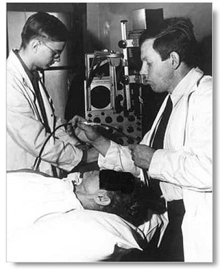
Wild and Reid, early 1950s.
|
Sonograms can be done abdominally or transvaginally. For pregnant women, abdominal ultrasounds are much more common. Transvaginal and transrectal ultrasounds are otherwise options for women who aren’t expecting since the images created are much clearer and precise. However, these kinds of ultrasounds can be used early on in the pregnancy if more detailed imaging is needed.
When should you get an ultrasound
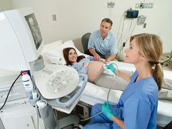
Sonography is able to view the baby as early as 20 weeks, the time period where the mother feels “quickening” or the first movement. The heart beat can be detected and the placement of both the placenta and the fetus can be determined. The same goes for the baby’s gender; it can verified at more or less 20 weeks.
Ultrasounds are used to detect:
Sonographers are very safe and are important prenatal diagnostic procedures to determine the health of the mother and child. If you are a parent expecting a child, be sure to get regular check-ups with your doctor to monitor how the pregnancy is doing.
Ultrasounds are used to detect:
- Presence of more than one fetus
- Due date and gestational age
- Baby’s health
- Location of the placenta
- Position of the fetus
- Expected weight of the fetus
Sonographers are very safe and are important prenatal diagnostic procedures to determine the health of the mother and child. If you are a parent expecting a child, be sure to get regular check-ups with your doctor to monitor how the pregnancy is doing.
