Transvaginal ultrasounds are routine examinations performed to assess a woman’s obstetric and gynecologic health.
For sonographers who specialize in OB-GYNE sonography, these are the most common type of examination they will perform in their career. While there are different ways to assess the female reproductive system, transvaginal and transrectal procedures are clearer because of the proximity of the transducer to the organ to be imaged.
The procedure
A transvaginal procedure is performed with the patient in a lithotomy position or dorsal recumbent. In the former, the patient lies down with her lower limbs in stirrups. The latter is similar, but instead of stirrups, the lower limbs are bent at an angle while laying flat on the examination table or bed. The lithotomy position is more commonly used because of better visualization and angle of insertion of the transducer.
The machine
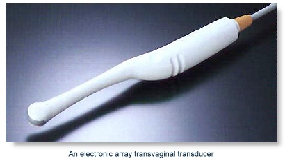
The transducer used in a transvaginal ultrasound is part of an elongated probe, roughly an inch or so in diameter. Because transvaginal ultrasounds are typically done to visualize the uterus, the probe is inserted until the cervix and the sonogram is started. The probe may be moved around the area for better visualization.
Like other sonography procedures, a transvaginal ultrasound also functions through the use of sound waves that return as electrical impulses which in turn create the images read by the machine. You can read more about ultrasound machine parts and function through the link.
Saline Infusion Sonohysterography (SIS)
The SIS is a special kind of transvaginal ultrasound where a volume of saline is introduced into the uterus. Saline is an IV fluid that contains 0.9 percent of sodium chloride, usually used in fluid resuscitation or electrolyte replacement. Saline is used in SIS in order to clearly see the endometrium (lining of the uterus) in the scan. After instilling the solution into the uterus, the sonogram proceeds as usual. Sonographers will usually be trained to perform this kind of procedure on-the-job, or at least be introduced to it in undergraduate clinical training.
Preparing the patient for a transvaginal ultrasound
During undergraduate training, sonographers learn about how to differentiate abnormal from normal results when giving ultrasounds. Abnormal results from a tranvaginal ultrasound usually identify problems such as birth defects (for prenatal transvaginal ultrasounds), cancer of the uterus, ovaries, vagina, cervix, and other similar structures, infections (including pelvic inflammatory disease [PID]), and structural abnormalities.
The CAAHEP has accredited more than 200 schools in the US that offer certificate, associate, or bachelor degrees in sonography. Enroll in a general sonography course and learn more about becoming a sonographer who specializes in obstetrics and gynecology.
The procedure
A transvaginal procedure is performed with the patient in a lithotomy position or dorsal recumbent. In the former, the patient lies down with her lower limbs in stirrups. The latter is similar, but instead of stirrups, the lower limbs are bent at an angle while laying flat on the examination table or bed. The lithotomy position is more commonly used because of better visualization and angle of insertion of the transducer.
The machine
The transducer used in a transvaginal ultrasound is part of an elongated probe, roughly an inch or so in diameter. Because transvaginal ultrasounds are typically done to visualize the uterus, the probe is inserted until the cervix and the sonogram is started. The probe may be moved around the area for better visualization.
Like other sonography procedures, a transvaginal ultrasound also functions through the use of sound waves that return as electrical impulses which in turn create the images read by the machine. You can read more about ultrasound machine parts and function through the link.
Saline Infusion Sonohysterography (SIS)
The SIS is a special kind of transvaginal ultrasound where a volume of saline is introduced into the uterus. Saline is an IV fluid that contains 0.9 percent of sodium chloride, usually used in fluid resuscitation or electrolyte replacement. Saline is used in SIS in order to clearly see the endometrium (lining of the uterus) in the scan. After instilling the solution into the uterus, the sonogram proceeds as usual. Sonographers will usually be trained to perform this kind of procedure on-the-job, or at least be introduced to it in undergraduate clinical training.
Preparing the patient for a transvaginal ultrasound
- Transvaginal ultrasounds are typically performed a week or more after menstruation.
- Sonographers should also instruct patients to empty their bladders before the procedure to prevent the organ from interfering with the view of the uterus and other supporting structures.
- Health education is something that is usually forgotten by health care providers, but it is one the most important duties of a sonographer. Teach the patient that a sonogram is typically done to detect abnormal growths such as polyps or a thickened endometrium, abnormal bleeding, ectopic pregnancies, and even certain types of infertility.
During undergraduate training, sonographers learn about how to differentiate abnormal from normal results when giving ultrasounds. Abnormal results from a tranvaginal ultrasound usually identify problems such as birth defects (for prenatal transvaginal ultrasounds), cancer of the uterus, ovaries, vagina, cervix, and other similar structures, infections (including pelvic inflammatory disease [PID]), and structural abnormalities.
The CAAHEP has accredited more than 200 schools in the US that offer certificate, associate, or bachelor degrees in sonography. Enroll in a general sonography course and learn more about becoming a sonographer who specializes in obstetrics and gynecology.
