|
Sonography has been around as early since the 1700, based on the discoveries by physicians on sound and echolocation. Today, it has evolved into one of the most popular diagnostic examinations performed in the hospital and diagnostic laboratories. Sonography as a diagnostic exam can be used for different parts of the body, from the abdomen to very small blood vessels. It is the mainstay in gynecology, serving as the gold standard for screening for numerous gynecologic conditions.
|
|
A little bit about sonography’s history
The use of a sonography as a diagnostic method was based on the concept of sound, first introduced in 1794 by Lazzaro Spallanzani, and how sound was used by animals with poor vision in order to move through their surroundings. The concept of sound travelling through empty space and bouncing off of solid structures was the focus when sonography was first introduced as a diagnostic procedure in 1877 by Pierre and Jacques Currie. The brothers used ultrasound probes that emitted and received sound waves using the piezoelectric effect.
It was in the late 1900s that sonography became more technologically advanced, using a sophisticated system that was capable of quality 3D – and eventually 4D – imaging. The sonograms to assist in medical procedures such as biopsies were also first introduced in the late 1900s. Today, sonography has become even more advanced, with the development of tablet-based sonography systems. More and more accredited diagnostic medical sonography schools are opening in the US, with more than 200 colleges currently accredited by the CAAHEP. Some of these schools even offer online classes.
Gynecologic sonographers
Gynecology is a branch of medicine that focuses on the female reproductive system. It usually goes hand in hand with obstetrics, a branch that deals with pregnancy, childbirth, and the postpartum period (perinatal approach). Sonography plays a big role in general gynecology, because surgical (invasive) procedures are often very risky for the female reproductive organs. Most women therefore prefer non-invasive diagnostic procedures, particularly sonography.
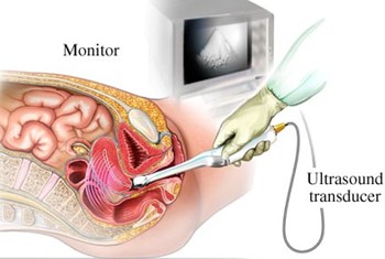
Gynecologic sonography can be performed abdominally, like a simple abdominal procedure, or transvaginally or transrectally. Trasvaginal and transrectalsonography is classified as an invasive version of this procedure because it involves the insertion of the probe into the vagina or rectal. These two procedures are much more accurate than performing sonography through the abdomen because the probe is placed closer to the uterus and other internal organs.
Screening for cancer
One of the most dangerous conditions that affect the female reproductive organs is cancer. Most women will only get a gynecologic ultrasound once they start experiencing signs and symptoms, usually pain or vaginal discharge. This is quite alarming because cancer can be caught early with the right diagnostic procedure, especially if the person gets regularly screened. But how often should a person get screened by an ultrasound technician?
Ultrasounds are being consistently used in diagnosing breast, cervical, and uterine cancer because the procedure is generally painless and poses no threat to immunocompromised patients. For cervical and uterine cancer, pap smears are the gold standard for detecting certain conditions. For breast cancers, ultrasounds are preferred over mammograms today because they don’t cause any pain, unlike the latter.
Gynecologic sonographers are one of the most in demand medical professions today. They can earn up to $100,000 annually depending on where in the US they are working. On average, sonographers earn an annual salary of $67,170 (according to the latest Bureau of Labor Statistics report). If you plan on choosing sonography as a career, consider gynecology as an option when choosing your ultrasound specialty.
The use of a sonography as a diagnostic method was based on the concept of sound, first introduced in 1794 by Lazzaro Spallanzani, and how sound was used by animals with poor vision in order to move through their surroundings. The concept of sound travelling through empty space and bouncing off of solid structures was the focus when sonography was first introduced as a diagnostic procedure in 1877 by Pierre and Jacques Currie. The brothers used ultrasound probes that emitted and received sound waves using the piezoelectric effect.
It was in the late 1900s that sonography became more technologically advanced, using a sophisticated system that was capable of quality 3D – and eventually 4D – imaging. The sonograms to assist in medical procedures such as biopsies were also first introduced in the late 1900s. Today, sonography has become even more advanced, with the development of tablet-based sonography systems. More and more accredited diagnostic medical sonography schools are opening in the US, with more than 200 colleges currently accredited by the CAAHEP. Some of these schools even offer online classes.
Gynecologic sonographers
Gynecology is a branch of medicine that focuses on the female reproductive system. It usually goes hand in hand with obstetrics, a branch that deals with pregnancy, childbirth, and the postpartum period (perinatal approach). Sonography plays a big role in general gynecology, because surgical (invasive) procedures are often very risky for the female reproductive organs. Most women therefore prefer non-invasive diagnostic procedures, particularly sonography.
Gynecologic sonography can be performed abdominally, like a simple abdominal procedure, or transvaginally or transrectally. Trasvaginal and transrectalsonography is classified as an invasive version of this procedure because it involves the insertion of the probe into the vagina or rectal. These two procedures are much more accurate than performing sonography through the abdomen because the probe is placed closer to the uterus and other internal organs.
Screening for cancer
One of the most dangerous conditions that affect the female reproductive organs is cancer. Most women will only get a gynecologic ultrasound once they start experiencing signs and symptoms, usually pain or vaginal discharge. This is quite alarming because cancer can be caught early with the right diagnostic procedure, especially if the person gets regularly screened. But how often should a person get screened by an ultrasound technician?
Ultrasounds are being consistently used in diagnosing breast, cervical, and uterine cancer because the procedure is generally painless and poses no threat to immunocompromised patients. For cervical and uterine cancer, pap smears are the gold standard for detecting certain conditions. For breast cancers, ultrasounds are preferred over mammograms today because they don’t cause any pain, unlike the latter.
Gynecologic sonographers are one of the most in demand medical professions today. They can earn up to $100,000 annually depending on where in the US they are working. On average, sonographers earn an annual salary of $67,170 (according to the latest Bureau of Labor Statistics report). If you plan on choosing sonography as a career, consider gynecology as an option when choosing your ultrasound specialty.
