|
Sonography is a popular diagnostic medical procedure that uses sound waves to create images of internal structures in the body. It is a preferred method of diagnosis for patients who are pregnant or have compromised immune systems because sound waves are safe and the procedure itself is performed non-invasively. As technological advancements were made in the 2000s, the procedure because highly specialized – able to produce three-dimensional images.
Accredited ultrasound technician programs offer training in 2D and 3D imaging, even 4D sonography, because their curricula include the latest skills and techniques. There are 3 online sonography schools that are accredited in the US, out of 200. Learn how to differentiate 2D and 3D imaging below. |
Training in diagnostic medical sonography
There are currently more than 200 schools in the US that offer the diagnostic medical sonography (DMS) program. These diagnostic medical sonography accredited schools (a list of available schools through the link) offer training in the latest skills and techniques in sonography, in both 2D and 3D imaging. Accredited schools have the best programs because their curricula have passed the nationwide standards set by the Commission on Accreditation of Allied Health Education Programs (CAAHEP).
Back to basics: The 2D ultrasound
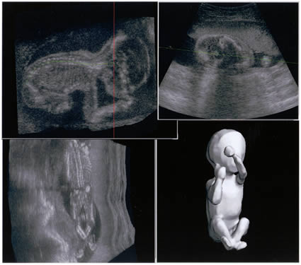
Before newer techniques were developed and machines were further advanced, two-dimensional images were the forte of a sonogram machine. Two-dimensional images simply mean a flat, black and white image of an internal structure, like a 2D cartoon on TV. This procedure is easier to perform (and generally more affordable) because the image produced is enough to get a preliminary diagnosis on the patient’s condition. More specialized tests are only performed after a preliminary diagnosis.
However, there are a lot of drawbacks to a 2D ultrasound. The image produced, while clear and can be used to make an initial diagnosis, is best used for the preliminary part of medical management. Most of the time, additional tests are needed in order to get better visualization of the affected area.
Three-dimensional imaging
The first 3D images were created in 1961 by Baum and Greenwood. They created the image by using a series of 2D images of the eye, reversed, on transparent plates. The images were compounded by stacking the plates with appropriate spacing, creating the three-dimensional image of the eye. Though this technique was successful in creating an anatomically correct 3D image, certain areas of the eye could not be seen clearly because superficial echoes from the other images when they were compounded.
Three-dimensional imaging can now be performed with modified sonogram machines. A 2D image is created by sending sound waves through the body in a straight line, which returns to the machine and creates the image. In 3D imaging, the sound waves are not sent in straight line, instead they are sent in angles. The waves reach the machine and are processed by an advanced program to create the image. This process was created in 1987 by von Ramm and Smith from Duke University.
Associated risks with sonography
According to research, there are no major risks associated with the ultrasound procedure. Sound waves, though sent in high frequency through the body, are perfectly safe, even for fetuses and immunocompromised patients. Ethical issues have been raised regarding “memento” sonograms, when parents get sonograms without a medical reason or indication – but generally, the procedure is quite safe and can be performed without affecting the mother, child, or patient.
There are currently more than 200 schools in the US that offer the diagnostic medical sonography (DMS) program. These diagnostic medical sonography accredited schools (a list of available schools through the link) offer training in the latest skills and techniques in sonography, in both 2D and 3D imaging. Accredited schools have the best programs because their curricula have passed the nationwide standards set by the Commission on Accreditation of Allied Health Education Programs (CAAHEP).
Back to basics: The 2D ultrasound
Before newer techniques were developed and machines were further advanced, two-dimensional images were the forte of a sonogram machine. Two-dimensional images simply mean a flat, black and white image of an internal structure, like a 2D cartoon on TV. This procedure is easier to perform (and generally more affordable) because the image produced is enough to get a preliminary diagnosis on the patient’s condition. More specialized tests are only performed after a preliminary diagnosis.
However, there are a lot of drawbacks to a 2D ultrasound. The image produced, while clear and can be used to make an initial diagnosis, is best used for the preliminary part of medical management. Most of the time, additional tests are needed in order to get better visualization of the affected area.
Three-dimensional imaging
The first 3D images were created in 1961 by Baum and Greenwood. They created the image by using a series of 2D images of the eye, reversed, on transparent plates. The images were compounded by stacking the plates with appropriate spacing, creating the three-dimensional image of the eye. Though this technique was successful in creating an anatomically correct 3D image, certain areas of the eye could not be seen clearly because superficial echoes from the other images when they were compounded.
Three-dimensional imaging can now be performed with modified sonogram machines. A 2D image is created by sending sound waves through the body in a straight line, which returns to the machine and creates the image. In 3D imaging, the sound waves are not sent in straight line, instead they are sent in angles. The waves reach the machine and are processed by an advanced program to create the image. This process was created in 1987 by von Ramm and Smith from Duke University.
Associated risks with sonography
According to research, there are no major risks associated with the ultrasound procedure. Sound waves, though sent in high frequency through the body, are perfectly safe, even for fetuses and immunocompromised patients. Ethical issues have been raised regarding “memento” sonograms, when parents get sonograms without a medical reason or indication – but generally, the procedure is quite safe and can be performed without affecting the mother, child, or patient.
