Ultrasound or sonography is a commonly non-invasive procedure performed most often for obstetric, gynecologic, and abdominal cases. However, as sonograms became more specialized, some procedures could also be performed invasively, such as intravascular ultrasound (IVUS). There are non-invasive alternatives to these special procedures but these have shown to have better accuracy than performing Doppler ultrasounds.
Because IVUS and other similar procedures are used in the vessels, the use of contrast is a practice that is done to guide the insertion of the catheter. In other cases, contrast may be used to simply improve the image quality of non-invasive ultrasound.
Because IVUS and other similar procedures are used in the vessels, the use of contrast is a practice that is done to guide the insertion of the catheter. In other cases, contrast may be used to simply improve the image quality of non-invasive ultrasound.
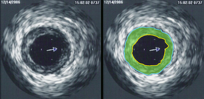
Intravascular ultrasound image of a coronary artery (left), with color-coding on the right, delineating the lumen (yellow), external elastic membrane (blue) and the atherosclerotic plaque burden (green). The percentage stenosis is defined as the area of the lumen (yellow) divided by the area of the external elastic membrane (blue) times 100. As the plaque burden increases, the lumen size will decrease and the degree of stenosis will increase.
The low-contrast problem
One of the major difficulties that non-invasive procedures have is a low-contrast between blood and surrounding tissues. This makes it difficult to discern different structures and tissues in the image generated by the sonogram. Doppler sonography can only be used in detecting blood flow in vessels that are of a certain size – any smaller than that size and the results have lower accuracy. This is the same reason why procedures like angiography or other forms of invasive and non-invasive diagnostic procedures use contrast to help guide the catheter or to position a patient.
UCA: ultrasound contrast agents
UCAs have actually been used for a long time, with the first reports of its use in 1968 but Gramiak and Shah in echocardiography. Because these UCAs were very rudimentary, there were simply room air bubbles that had no protective covering and disappeared within a few seconds after administration. Modern-day UCAs are much more stable, made up of gases with low-diffusion rates like nitrogen or perfluorocarbon that is further stabilized by coating the UCA with biodegradable material (albumin, phospholipids).
Ultrasound education
There are more than 200 schools in the US in 2014 that are accredited institutions. They offer quality training programs whose curricula meet the standards set throughout the nation. Accredited diagnostic medical sonography schools in Florida are some of the best in the country, offering the best training in Miami. If you study in these accredited programs, you will able to pass the ARDMS exam with flying colors and learn more about UCAs and sonography developments.
The use of these UCAs can help distinguish the blood vessel and body cavity walls from blood flow and other organs - all because of high-contrast. UCAs can be used adjunct with invasive or non-invasive ultrasound methods, with no harm coming to the patient (unless he or she is allergic to iodine-based dyes).
The use of these UCAs can help distinguish the blood vessel and body cavity walls from blood flow and other organs - all because of high-contrast. UCAs can be used adjunct with invasive or non-invasive ultrasound methods, with no harm coming to the patient (unless he or she is allergic to iodine-based dyes).
