Congenital heart defects are quite serious medical conditions that require intensive management once diagnosed. For some conditions, the signs and symptoms only manifest as the child gets older and becomes more active – typically during the toddler years. However, there are certain heart defects that can lead to death within minutes to hours of the birth if not diagnosed and managed early. Sonography is one of the most overlooked procedures than can help with different diagnoses.
The role of sonography in diagnosis
Sonography is one of the most commonly used diagnostic procedures today. It is safer than other diagnostic procedures such as MRIs and X-rays because it doesn’t use radiation, allowing pregnant mothers, children, and immunocompromised people to get an ultrasound without any worries. Sonography is commonly used on the abdomen and reproductive organs but can be used for other body systems as well.
Instead of radiation waves that react with certain dyes injected in the blood, sonography just sends out sound waves that pass through the body and back to the sonogram machine. These sound waves bounce off from solid structures and create images as they return to the machine at certain intervals.
Instead of radiation waves that react with certain dyes injected in the blood, sonography just sends out sound waves that pass through the body and back to the sonogram machine. These sound waves bounce off from solid structures and create images as they return to the machine at certain intervals.
Congenital defects in fetuses
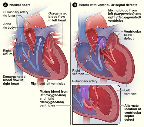
Congenital heart disease (CHD) is among the most common causes of mortality in infants and children. It is also the leading organ-specific congenital defect in the United States, with more than half of children with CHD born to mothers without any previously known risk factors, both hereditary and non-hereditary. With these statistics, there is a need for an effective diagnostic procedure that will be able to detect cardiac defects in fetuses.
Updates in sonography
New sonography techniques have been developed over the last several years, making real-time visualization of the body’s internal structures possible. One of these new techniques is called B-flow ultrasound – a procedure that is able to give real-time visualization of the blood flow in certain areas of the body. It was first used as a cardiac and vascular procedure but studies have shown that it is excellent for diagnosing fetal health conditions.
Standards in diagnosis
While 2D echocardiograms are still the gold standard for diagnosing fetal health defects, B-flow sonography is a unique technique that can be used in its place. It doesn’t rely on the use of Doppler and is much easier to use. B-flow sonography uses digitally-encoded technology to suppress tissue clutter in the images and improve the sensitivity of the procedure. However, the procedure cannot be used if the fetus is in a certain position and if the fetal heart rate is too fast. Unlike a 2D-echo, the B-flow technique can visualize smaller, peripheral vessels with low velocity, making the results more accurate and helping get a complete, concrete diagnosis compared to other procedures.
Studying updated sonography techniques
There are 211 schools that have sonography programs in the US in 2014, with only three with sonography programs online. There 211 programs are all accredited by the CAAHEP, meaning that their curricula meets the standards set all over the nation. CAAHEP-accredited programs are sure to have the latest sonography techniques and updates compared to non-accredited programs. You can find more details on sonography programs and how to choose the right one for you in Lisa’s articles.
B-flow sonography is not the only new technique in sonography. If you study and train in a post-graduate course, there are many more techniques and updates that you can learn that can make your skills very competitive – especially if you’re applying to a job. These updated techniques are not necessarily replacing the gold standards for diagnosis certain conditions but they can help improve the statistics for early diagnosis and management of congenital defects in foetuses.
B-flow sonography is not the only new technique in sonography. If you study and train in a post-graduate course, there are many more techniques and updates that you can learn that can make your skills very competitive – especially if you’re applying to a job. These updated techniques are not necessarily replacing the gold standards for diagnosis certain conditions but they can help improve the statistics for early diagnosis and management of congenital defects in foetuses.
