Diagnostics is a very important part of healthcare because it detects possible health maladies that would’ve otherwise been left unmanaged. Sonography is a diagnostic procedure that uses sound waves to create images of structures within the body, from organs, bones, and even tumors and growths. A healthcare provider tasked with operating a sonogram is a sonographer; a sonographer is trained how to use the sonogram, particularly how to manipulate the transducer (in order to get the desired image) and analyze the images the machine produces.
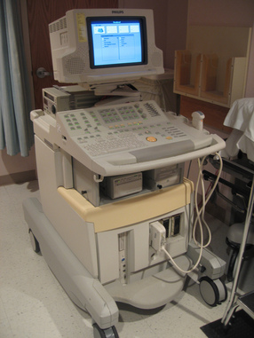
The sonogram
|
The sonogram is a machine that has two basic parts, the monitor and the transducer. The transducer is a small, hand-held apparatus that sends out high-frequency sound waves that pass through the body. When these sound-waves bounce off at intervals at the structures in the body, they create images on the monitor. The sonographer then prints out the images and analyzes normal from abnormal findings. This procedure is also referred to as an ultrasound. For more information on the procedure click here.
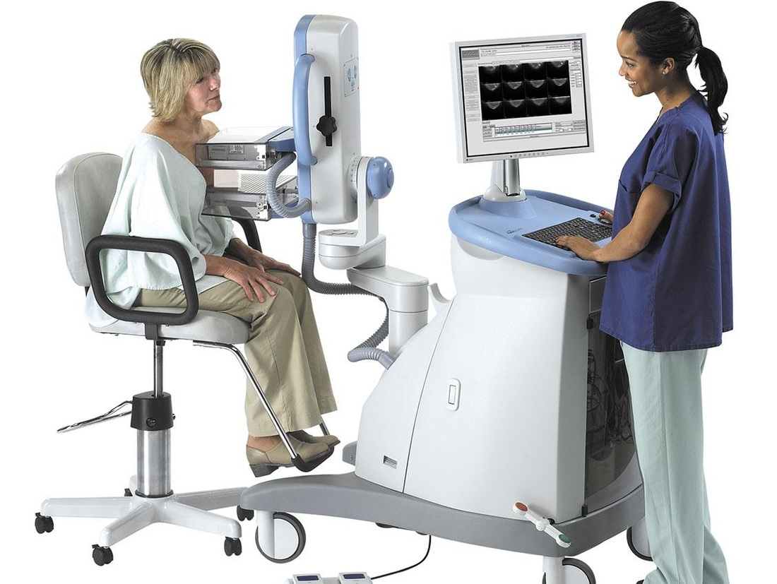
Screening for breast tumors
With new discoveries in healthcare in 2014, sonography is no longer being confined to obstetrics and gynecologic purposes. They can be used to screen for anomalies such as tumors in areas that don’t normal undergo ultrasound – particularly the breasts. The accepted methods for screening for breast cancer are mammography and palpation (through a breast exam). However, reports of false-negatives and differences among reported series have caused a decrease in the effectiveness of these screening procedures.
Since breasts are different in size, with some breasts denser than the others, the sensitivity of mammography for screening varies per individual. Through studies done on mammography, sensitivity for the procedure is lower in radiographically dense breasts – typically seen younger, pre-menopausal women. The test is significantly more sensitive in women older than 50 years old. This is troubling because the results from a mammography can only be considered truly significant among older women, causing poor diagnosis of possible breast problems in younger women. Mammography and sonography
The results of a recent study on women with breast lesions in 2012 show that mammography sensitivity was 81.71% compared to the 95.53% of sonography. Positive predictive values (PPVs) were 76.72% and 74.13% respectively. Negative predictive values were likewise 88.83% and 96.84% respectively. These statistics are significant for both procedures, showing that mammography is still a golden standard for breast cancer screening, sonography comes at a very close second, even showing to be more sensitive in detection than the former.
|
|
Breast sonography
With the positive results from the study, breast sonography is a relatively new avenue that sonographers can enter or consider when looking for the right specialty for them. The American Registry of Radiologic Technologists (ARRT) gives certification for the specialty of breast sonography. You can read more about certification and specialties in Lisa’s articles through the link.
