|
For sonographers who work in obstetrics and gynecology, confirming a pregnancy is one of the most common indications for uterine ultrasound. But how exactly is a pregnancy confirmed? The most basic thing is to identify the gestational sac in the sonogram, but there are many ins and outs when performing the procedure itself. A sonographer learns OB-GYNE sonography through a general sonography program, the most common program offered by educational institutions in the US.
|
Education in Sonography
Each year, the Commission on Accreditation of Allied Health Education Programs (CAAHEP) evaluates schools and their sonography programs. There are currently more than 200 schools in the US that have been granted accreditation by CAAHEP. There are sonography schools in almost every state, especially the most populous ones.
The third most populous state in the country is Chicago, and is home to two accredited educational institutions. Because of the number of people living in Chicago, the demand for sonographers has been steadily increasing. Because of this demand, the number of sonography programs in Chicago can be expected to increase as well.
Definitive diagnosis
When a woman wants to find out if he or she is pregnant, there are two pathways that they usually take – take a home pregnancy test (which is used to measure levels of hCG in the blood) followed by an ultrasound or bypass the first step and proceed to a diagnostic procedure. The sonogram is still the gold standard for detecting a normal pregnancy, because while human chorionic gonadotropin (hCG) is produced by a fertilized egg, high levels can also be indicative of other conditions.
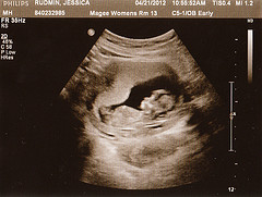
In a sonogram as early as three weeks, a gestational sac can be seen. In a normal pregnancy, the sac is implanted along the walls of the uterus, containing a yolk sac or an embryo/fetus. The placenta can also be visualized. The sonogram will show a small, intrauterine fluid collection within the sac if the sonogram is performed early on in the pregnancy.
In an abnormal pregnancy, there is usually high hCG levels without visual confirmation from the uterus. Additional tests are performed, and usually a sonogram is also used to detect if the embryo implanted somewhere else in the gestational tract (most likely the fallopian tubes). In special cases, the embryo can even implant outside of the lower pelvic area.
The safety of transvaginal scans
Most pregnant women are afraid to get transvaginal or transrectal sonograms. However, a transvaginal sonogram is quite safe for the mother and child as long as the mother remains calm throughout the procedure. The transducer is a long thin wand only placed just below the cervix, so no harm is done to the fetus.
However, this kind of exam can be contraindicated in pregnancies where the women has a previous history of bleeding or an incompetent cervix. Otherwise, the procedure is quite safe for women with normal pregnancies. This kind of sonogram is actually much more accurate than an abdominal one, because of the proximity of the transducer to the uterus.
Sonographers should be able to prepare their patients well for a variety of procedures. Health education is very important, not confined to simply teaching them about the sonogram itself but for possible changes in their medical care and management depending on the results.
Each year, the Commission on Accreditation of Allied Health Education Programs (CAAHEP) evaluates schools and their sonography programs. There are currently more than 200 schools in the US that have been granted accreditation by CAAHEP. There are sonography schools in almost every state, especially the most populous ones.
The third most populous state in the country is Chicago, and is home to two accredited educational institutions. Because of the number of people living in Chicago, the demand for sonographers has been steadily increasing. Because of this demand, the number of sonography programs in Chicago can be expected to increase as well.
Definitive diagnosis
When a woman wants to find out if he or she is pregnant, there are two pathways that they usually take – take a home pregnancy test (which is used to measure levels of hCG in the blood) followed by an ultrasound or bypass the first step and proceed to a diagnostic procedure. The sonogram is still the gold standard for detecting a normal pregnancy, because while human chorionic gonadotropin (hCG) is produced by a fertilized egg, high levels can also be indicative of other conditions.
In a sonogram as early as three weeks, a gestational sac can be seen. In a normal pregnancy, the sac is implanted along the walls of the uterus, containing a yolk sac or an embryo/fetus. The placenta can also be visualized. The sonogram will show a small, intrauterine fluid collection within the sac if the sonogram is performed early on in the pregnancy.
In an abnormal pregnancy, there is usually high hCG levels without visual confirmation from the uterus. Additional tests are performed, and usually a sonogram is also used to detect if the embryo implanted somewhere else in the gestational tract (most likely the fallopian tubes). In special cases, the embryo can even implant outside of the lower pelvic area.
The safety of transvaginal scans
Most pregnant women are afraid to get transvaginal or transrectal sonograms. However, a transvaginal sonogram is quite safe for the mother and child as long as the mother remains calm throughout the procedure. The transducer is a long thin wand only placed just below the cervix, so no harm is done to the fetus.
However, this kind of exam can be contraindicated in pregnancies where the women has a previous history of bleeding or an incompetent cervix. Otherwise, the procedure is quite safe for women with normal pregnancies. This kind of sonogram is actually much more accurate than an abdominal one, because of the proximity of the transducer to the uterus.
Sonographers should be able to prepare their patients well for a variety of procedures. Health education is very important, not confined to simply teaching them about the sonogram itself but for possible changes in their medical care and management depending on the results.
