Sonography has come a long way from prenatal diagnostic examinations. The procedure is no longer confined as purely a diagnostic procedure; it is being used in real-time management and treatment. Sonography is used in surgery, assessment and treatment of trauma patients, and now in radiotherapy. The popularity of sonography comes from it “safe” characteristic, free from any radiation and emissions that can harm patients who are immunocompromised and/or pregnant.
Thyroid Cancer
Abnormal growths in the thyroid are called thyroid nodules, present in about 4 to 7 percent of the population. Out of 30 to 50 percent of autopsy cases in 2014, only 5 percent of these nodules are biopsied and found to be malignant (cancerous). Thyroid cancer is dangerous because the thyroid is a gland that stimulates the release of certain hormones. Hormones are responsible for maintaining function and balance in the body, and if the thyroid is unable to control them, it can cause a variety of crippling signs and symptoms.
Common signs and symptoms of thyroid cancer include:
Common signs and symptoms of thyroid cancer include:
- Swelling of the neck
- Pain in the neck and ears
- Difficulty swallowing
- Difficulty swallowing
- Difficulty breathing / wheezing
- Hoarse voice
- Frequent cough without a cold
Sonography and Sclerotherapy
One of the most common methods in treating thyroid cancer is through sclerotherapy. Sclerotherapy uses sclerosing agents like ethanol and tetracycline which are injected into the nodule. This kind of therapy is also used for treating varicose veins, to help reduce inflammation of the vessels. However, before sclerosing can be done, aspiration is usually performed first. Fine-needle aspiration biopsy (FNAB) is used to take a sample of the fluid in the nodule, as well as to aspirate it and reduce the nodule’s size.
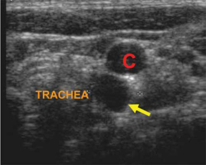
Because both of the methods involve puncturing the nodule, it is difficult without proper visualization. Invasive procedures can be quite risky, which is why physicians prefer to aspirate and manage the nodule with sclerotherapy. Sonography plays an important part because it is used to guide the needle as it takes a sample of fluid from the nodule as well as during the injection of the sclerosing agents (more information through the link). Nodules are typically filled with fluid, which can be detected by sonography in real time as the needle aspirates the fluid.
Because both of the methods involve puncturing the nodule, it is difficult without proper visualization. Invasive procedures can be quite risky, which is why physicians prefer to aspirate and manage the nodule with sclerotherapy. Sonography plays an important part because it is used to guide the needle as it takes a sample of fluid from the nodule as well as during the injection of the sclerosing agents (more information through the link). Nodules are typically filled with fluid, which can be detected by sonography in real time as the needle aspirates the fluid.
Studies about Sonography and Thyroid Disease
With the introduction of sonography in the management of thyroid problems, it has helped in diagnosis and treatment of thyroid cysts. While majority of cysts found in the thyroid are not malignant, sonography has helped established a gold standard for both preoperative diagnosis and treatment for them. You learn more about sonography in FAQs for sonography students through the link. If you want to study sonography, check degrees through this link http://www.ultrasoundtechniciancenter.org/category/search-by-degree.
These positive outcomes regarding the use of sonography have reduced the need for unnecessary thyroid surgery for ENT patients; a feat for a procedure that has been overlooked as a diagnostic tool for OB-GYNE and abdominal cases.
These positive outcomes regarding the use of sonography have reduced the need for unnecessary thyroid surgery for ENT patients; a feat for a procedure that has been overlooked as a diagnostic tool for OB-GYNE and abdominal cases.
