How can sonography help in weaning patients from mechanical ventilation? Learn more about this simple procedure today.
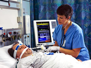
Mechanical ventilation is a form of management for patients experiencing respiratory failure, or have severe respiratory problems that impair their ability to breathe without assistance or spontaneously. While being mechanically ventilated may seem scary, it is an important lifesaving procedure that allows a patient to recover until he or she can breathe on his or her own. However, a problem with ventilation is difficulty weaning the patient from it. Patients are either weaned to early or too late – this is where sonography can help.
Diagnosis is the first step
The first step before any medical management is done is assessment. Before a patient can be weaned off from the ventilator, assessing his or her ability to spontaneous breathe must be done. For example, a patient has a rupture in the lung tissue that impairs his or her ability to respire so he or she was placed under mechanical ventilation. Once the tissue healed, the patient was prepared to be weaned from the machine. But how do you know that the patient is ready?
Most of the time, trial and error is the key. The settings are lowered and the patient is observed if he or she can withstand them. However, specific assessment as to whether a person can be weaned is difficult. One of the problems associated with difficulty weaning is a problem with the diaphragm. The diaphragm is responsible for allowing the lungs to expand by contracting, and compressing the lungs during exhalation by relaxing. If the diaphragm is too weak to support normal respiration, it can cause weaning problems.
An ultrasound of the abdomen
In order to assess if there is problem with the muscles of the diaphragm, B-mode sonography can be used. The procedure was done by placing the transducer perpendicular to the eight or ninth ICS (intercostal space), between the anterior axillary and midaxillary lines. Each frozen B-mode image is analyzed for the measurement of diaphragmatic thickness. This procedure is performed twice, once before the patient is placed on the ventilator for a comparison, and the other before weaning. If there is significant atrophy (loss of muscle), the patient is most likely unable to support spontaneous respiration.
The use of a sonogram is quite ideal because it is highly feasible, safe, and repeatable among different patients. It also makes weaning a patient from the ventilator much easier because there is a visual representation of his or her ability to breathe normally. You can inquire about this procedure in a continuing education class.
Learn how to become an ultrasound technician in 2016.
