Sonography is a diagnostic procedure that uses sound waves to create images of structures inside the human body. It is preferred to procedures such as MRIs, x-rays, and surgical exploration because it is safe as well as (generally) non-invasive. Compared to other procedures, it is quite cost-effective and risk-free, making it ideal for a variety of patients and cases. But how was ultrasound first invented? What was it first used for?
Non-audible sound and Echolocation
|
It was in the 1700s when Lazzaro Spallanzani, an Italian physiologist, conducted experiments that revealed the existence non-audible sound used in spatial orientation. He primarily cited bats who are blind but could navigate around different obstacles by creating sound from their mouths. He proved this by conducting experiments where bats were unable to avoid obstacles when their mouths we covered. This was termed “Spallanzani’s Bat Problem”.
It was Spallanzani’s works on sound that served as the basis for sonography – or ultrasound and other similar procedures. The non-audible sound that he hypothesized is today known as high-frequency sound waves that cannot be heard by the human ear. At whatever frequency, sound has one major characteristic; it is able to bounce off of solid structures such as tissues and bone. This is the same principle that is used in modern sonography. Learn about history of ultrasound from this website. |

|
The Piezo-electric effect
The development of the ultrasound transducer was made possible by the discoveries of the Currie brothers in 1880. They discovered that when electricity travels through certain crystals, they vibrate at a high frequency – creating a phenomenon they coined as the “Piezo-electric effect”. Today, ultrasound machines and their transducers are outfitted with thin sheets of crystal which vibrate and are responsible for creating the sound waves that pass through the body. During the same year, Galton was able to create a transducer using mechanical vibration of a sheet of quartz.
Medical ultrasound
By the 1900s, ultrasound was being tested in the medical field, setting out to create images of the inside of the human body without surgical intervention. Karl Dussik was a neurologist in 1942 that first attempted to use sound waves as a medical diagnostic tool. He used the machine to attempt to locate brain tumors and normal anatomical structures. This attempt was mostly unsuccessful because the sound waves were unable to penetrate the osseous layer of the skull.
The first, and one of the most important, papers published on medical sonography was by Donald and Brown in 1956. Donald had initially used sonography for anatomical measurements of fetal specimens. The one-dimensional equipment he used was further refined by Brown, creating the first 2D (two-dimensional scanner), called a “two-dimensional compound scanner”.
Commercially, sonogram machines were introduced and gained popularity in the 1960s – when B-mode scanning was developed. B-mode stands for brightness mode, a kind of sonography where a linear array was used to create 2D images. This mode is more commonly known as 2D scanning today and is still used for a variety of cases. Today, there are A, C, M, and Doppler modes used in medical sonography – all with different characteristics and uses.
|
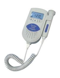
Using a Doppler
Doppler ultrasound is one of the most popular kinds of the procedure, because of its ability to take images of moving substances – particular blood flow through vessels. Dopplers are commonly used in obstetrics and gynecology for listening to fetal heartbeats and in vascular sonography, especially if an obstruction to a vessel is being considered. Sonography has come a long way since it was first developed as early as the 1700s. Today, it is an institution all on its own, with a reputation for being a specialized diagnostic procedure that has little to no risks and is very affordable and available. |
|
